Common Name
Deepwater Goby
Year Described
Ginsburg, 1953
Identification
Dorsal Fin: VII, 9
Anal Fin: 9
Pectoral Fin: 16
Caudal Fin: 17 segmented rays
Vertebrae: 11+16= 27 (total)
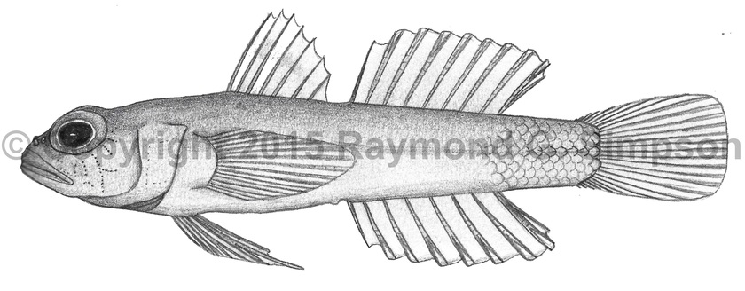
Body stout anteriorly and tapering posteriorly. Eye relatively large. Dorsal fin with anterior two spines not elongate and last two spines more spaced than the first five. Pelvic fin rays branched with the last relatively short. Pelvic fins not fused. One anal-fin pterygiophore anterior to the first haemal spine. Papillae rows 5i and 5s are separate. Cephalic lateralis pores absent. Body scaled anteriorly to about middle of second dorsal fin (12 rows). Belly unscaled. Basicaudal scales present.
Color
Color in preservative noted as pale yellow. Hastings & Bortone (1981) describe a specimen from off the Yucatan that they identify as this species that was pale yellow overall with a brighter yellow head and no markings.
Size
Only known specimen 30.7mm SL.
Habitat
Taken in deep water (154m).
Range
Known from a single specimen off the Yucatan Peninsula, Mexico.
References
Ginsburg, I. 1953. Ten new American gobioid fishes in the United States National Museum, including additions to a revision of Gobionellus. Journal of the Washington Academy of Sciences v. 43 (no. 1): 18-26.
Hastings, P.A. & S.A. Bortone. 1981. Chriolepis vespa, a new species of gobiid fish from the northeastern Gulf of Mexico. Proceedings of the Biological Society of Washington v. 94 (no. 2): 427-436.
Tornabene, L., J.L. Van Tassell, R.G. Gilmore, D.R. Robertson, F. Young, & C.C. Baldwin. 2016. Molecular phylogeny, analysis of character evolution, and submersible collections enable a new classification of a diverse group of gobies (Teleostei: Gobiidae: Nes subgroup), including nine new species and four new genera. Zoological Journal of the Linnean Society.
Other Notes
This species was placed in Varicus by Tornabene et al. (2016) based on the single anal pterygiophore placement (vs two in Pinnichthys), scaled rear body (versus fuly scaled or naked), and low meristics, but differs from the “typical” Varicus by papillae pattern (separate) and having branched pelvic rays. More study is needed when more specimens can be examined.